Home > For Her Wellness > Normal Service

Normal Services

Anomaly Scan

Anomaly Scan

An anomaly scan or a mid-pregnancy ultrasound scan is a detailed examination performed around 18-22 weeks of pregnancy. Its primary purpose is to assess the baby’s growth and development, as well as to detect any structural abnormalities. During the scan, a trained sonographer examines various aspects, such as the baby’s head, brain, spine, heart, kidneys, limbs and umbilical cord. They also check the placenta and amniotic fluid levels. The scan provides crucial information to parents and doctors, helping to ensure the baby’s health and well-being throughout pregnancy and into birth, guiding any necessary medical interventions or preparations.

Growth Scan

Growth Scan with Doppler Assessment

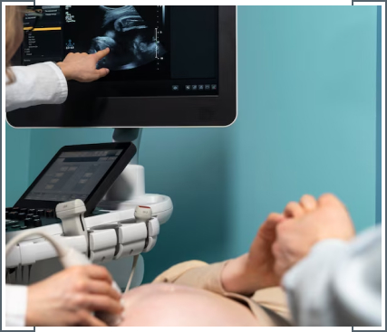
A growth scan with Doppler assessment is a specialized ultrasound procedure performed during pregnancy to evaluate the baby’s growth and assess blood flow within the umbilical cord and various fetal vessels. The scan measures the baby’s size, particularly focusing on head and abdominal circumference and femur length, to monitor growth patterns and detect any deviations from expected norms.

Doppler ultrasound is used to examine blood flow in the umbilical artery, middle cerebral artery and other vessels, providing insights into placental function and fetal well-being. This comprehensive assessment helps doctors monitor fetal development, identify potential issues early and make informed decisions regarding the management of pregnancy.

Growth Scan
Cervical Length

Cervical Length Assessment

Cervical length assessment is a crucial tool in obstetrics that is used to predict the risk of preterm birth. Usually measured via ultrasound, it evaluates the length of the cervix, which shortens as labour approaches. A shorter cervix early in pregnancy increases the likelihood of premature delivery. This non-invasive screening helps doctors identify high-risk pregnancies early, allowing for timely interventions to reduce the risk of preterm birth and improve maternal and neonatal outcomes. Regular monitoring throughout pregnancy ensures proactive management for optimal maternal and fetal health.

Early Pregnancy

Early Pregnancy Scan

An early pregnancy scan, usually performed between 6 to 10 weeks gestation, is a vital diagnostic tool for expecting mothers. Using ultrasound technology confirms pregnancy viability by detecting the fetal heartbeat, assessing gestational age and identifying multiple pregnancies or abnormalities. This scan provides reassurance to parents, ensures proper prenatal care planning and helps detect any potential issues early on. Early detection allows doctors to offer timely interventions or support as needed, fostering a smoother pregnancy journey and enhancing the overall health outcomes for both mother and baby.
Early Pregnancy
Dating Scan

Dating Scan

A dating scan, usually performed around 8-14 weeks of pregnancy, accurately determines gestational age and estimated due date. Using ultrasound technology, it measures the size of the fetus, assesses its development and confirms the presence of a heartbeat. This scan is crucial for establishing a timeline of the pregnancy, ensuring proper prenatal care and identifying any potential concerns early on. It provides expectant parents with essential information about their baby’s growth and development, offering reassurance and guiding doctors in creating care plans for a healthy pregnancy journey.
Viability Scan

Viability Scan

A viability scan, generally conducted around 6-10 weeks of pregnancy, confirms the presence of an intrauterine pregnancy and assesses fetal viability. Using ultrasound, it checks for a fetal heartbeat, measures the gestational sac and evaluates early fetal development. This scan is essential for reassuring expectant parents, especially those experiencing symptoms like bleeding or pain, by confirming the pregnancy’s progress and ruling out miscarriage or ectopic pregnancy. Early detection of viability issues allows doctors to offer appropriate guidance and support.
Viability Scan
Fetal Echocardiography

Fetal Echocardiography Scan

Fetal echocardiography is a specialized ultrasound procedure performed during pregnancy to evaluate the developing baby’s heart. It gets conducted between 18-24 weeks gestation, it assesses the structure and function of the fetal heart, detecting any congenital heart defects or abnormalities. This non-invasive imaging technique provides detailed information to doctors, enabling early diagnosis and management of cardiac conditions before birth.
NIPT-img

NIPT

Non-Invasive Prenatal Testing (NIPT) is an advanced screening method used during pregnancy to assess the risk of chromosomal abnormalities like Down syndrome (Trisomy 21), Trisomy 18, and Trisomy 13. It involves analyzing cell-free fetal DNA circulating in the mother’s bloodstream, obtained through a simple blood draw. NIPT offers high accuracy in detecting these conditions, with a lower risk of miscarriage compared to invasive procedures like amniocentesis or chorionic villus sampling. Results are available within a few days to weeks, providing expectant parents with valuable information for decision-making regarding further diagnostic testing and pregnancy management.
NIPT-img
Ultrasound assessment

Ultrasound assessment of pregnancies with fibroids

Ultrasound assessment of pregnancies with fibroids involves evaluating the presence, size, location and impact of uterine fibroids on the developing fetus and pregnancy. Fibroids are non-cancerous growths in the uterus that can vary in size and position, potentially affecting pregnancy outcomes. Ultrasound helps monitor fibroid growth, assesses its potential to cause complications such as miscarriage, preterm labour or abnormal fetal position and guides management strategies. Regular monitoring allows doctors to adjust treatment plans as needed to optimize maternal and fetal health throughout pregnancy.
Anomaly Scan

3D/4D Ultrasound

3D/4D ultrasound technology provides a detailed, three-dimensional view of the fetus during pregnancy, enhancing visualization beyond traditional 2D scans. It captures realistic images and movements, offering expectant parents a clearer perspective of their baby’s facial features, limbs and overall development. This advanced imaging can aid in detecting structural abnormalities and assessing fetal growth with greater precision. While primarily used for bonding and emotional connection, 3D/4D ultrasound may complement medical assessments in specific cases. However, it’s important to note that this technology is optional and should be used judiciously under the guidance of doctors to ensure safe and appropriate prenatal care.
Anomaly Scan
Müllerian anomalies

Müllerian anomalies ultrasound for multiple pregnancies and monochorionic twin pregnancy complications

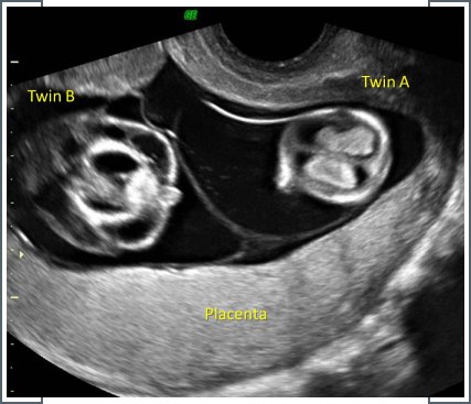
Ultrasound for multiple pregnancies, particularly in cases of monochorionic twins or pregnancies complicated by Müllerian anomalies, is essential for assessing fetal development and monitoring potential complications. Müllerian anomalies involve structural abnormalities in the uterus, impacting fertility and pregnancy outcomes. In monochorionic twins who share a placenta, ultrasound helps detect complications like twin-to-twin transfusion syndrome, where blood flow between twins is imbalanced. Early detection through ultrasound enables timely intervention, ensuring proper management to mitigate risks such as preterm birth or developmental issues.